Manual of Surgery - LightNovelsOnl.com
You're reading novel online at LightNovelsOnl.com. Please use the follow button to get notifications about your favorite novels and its latest chapters so you can come back anytime and won't miss anything.
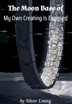
[Ill.u.s.tration: FIG. 60.--Section through Hip-Joint to show epiphyses at upper end of femur, and their relation to the joint.
_a_, Epiphysis of head.
_b_, Epiphysis of great trochanter.
_c_, Epiphysis of small trochanter.
_d_, Capsular ligaments.
(After Poland.)]
FRACTURE OF THE NECK
It has long been customary to divide fractures of the neck of the femur into two groups--"intra-" and "extra-capsular"; but as in a considerable proportion of cases the line of fracture falls partly within and partly without the capsule, this cla.s.sification is wanting in accuracy. It is more correct to divide these fractures into (1) those occurring _through the narrow part of the neck_, which are nearly always purely intra-capsular; and (2) those occurring _through the base of the neck_ in which the line of fracture lies inside the capsule in front, but outside of it behind.
It is of considerable importance to distinguish between fractures in these two positions. The first group occurs almost exclusively in old persons as a result of slight forms of indirect violence, and it is liable, on account of the feeble vascular supply to the upper fragment, to be followed by absorption of the neck, which delays or may even entirely prevent union (Fig. 61). The second group usually occurs in robust adults, and results from severe forms of violence applied to the trochanter. In this group firm osseous union usually takes place.
[Ill.u.s.tration: FIG. 61.--Fracture through Narrow Part of Neck of Femur on section. The Neck of the bone has undergone absorption.]
#Fracture of the Narrow Part of the Neck# or #Intra-capsular Fracture#.--This fracture is most frequently met with in elderly persons, especially women, and is usually produced by comparatively slight forms of indirect violence--such, for example, as result from the foot catching on the edge of a carpet, a stumble in walking, or missing a step in going downstairs.
The line of fracture, which is usually transverse but may be oblique or irregular, lies for the most part within the capsule, and the posterior part of the neck is more comminuted than the anterior. The distal fragment, which includes the base of the neck, the trochanters, and the shaft, is usually displaced upward and rotated laterally. If the periosteum and the retinacular ligaments remain intact, displacement is prevented and union favoured.
Impaction is less common than in fracture through the base of the neck; it usually results from the patient falling on the trochanter, the distal fragment being driven as a wedge into the proximal (Fig.
62).
[Ill.u.s.tration: FIG. 62.--Impacted Fracture through Narrow Part of Neck of Femur.]
_Clinical Features._--In non-impacted cases the limb is at once rendered useless, and the patient is unable to rise. There is pain and tenderness in the region of the hip on making the slightest movement; and a specially tender spot may be localised, indicating the seat of fracture.
On placing the pelvis as square as possible, and comparing the measurements of the limbs from the anterior superior spine to the medial malleolus, shortening of the injured limb to the extent of from 1 to 3 inches may be found. On applying Nelaton's, Bryant's, or Chiene's test, the tip of the great trochanter will be found elevated.
It is also farther back and less prominent than normal.
The whole limb is usually everted to a greater or less degree, and is slightly abducted. In some cases, when the impaction is of the anterior portion of the neck, the limb is inverted. On comparing the ilio-tibial band of the fascia lata on the two sides, it is found to be relaxed on the side of the injury.
The violence being as a rule indirect, there is at first little or no discoloration in the vicinity of the hip, but this may appear a few days later.
Crepitus is not a constant sign, and should not be sought for, as the necessary manipulations are liable to disengage the fragments and to increase the deformity. For the same reason rotatory movements are to be avoided.
In all cases in which the diagnosis is uncertain, the patient should be put to bed, and treated as for a fracture. In the course of a few days it is nearly always possible to make an accurate diagnosis.
In examining an old person who has sustained an injury in the region of the hip, it should be borne in mind that the limb may be shortened and everted as a result of arthritis deformans, and that the symptoms of that disease may simulate those of fracture. In arthritis deformans, however, the ilio-tibial band of the fascia lata is not relaxed as it is in fracture.
[Ill.u.s.tration: FIG. 63. Fracture of Neck of Right Femur, showing shortening, abduction, and eversion of limb.]
In some cases, and particularly in those in which the periosteum of the neck and the retinacular ligaments remain intact, the shortening does not become apparent till a few days after the accident. As the other symptoms are correspondingly obscure, the condition is apt to be mistaken for a bruise. In all doubtful cases the part should be examined from day to day, and, if possible, the X-rays should be used.
In _impacted_ cases the signs of fracture are often obscure, and the patient may even be able to walk after the accident. The skin over the trochanter is generally discoloured from bruising. Eversion is usually present, but there may be little shortening. Crepitus is absent. In old people it is never advisable to undo impaction, as the interlocking of the bones favours the occurrence of osseous union.
_Prognosis._--A fracture of the neck of the femur in an old person is always attended with danger to life, a considerable proportion of the patients dying within a few weeks or months of the accident from causes a.s.sociated with it. In some cases the mental and physical shock so far diminishes the vitality of the patient that death ensues within a few days. It is possible that fat embolism may account for death in some of the more rapidly fatal cases. In others, the continued dorsal position induces hypostatic congestion of the lungs, or, owing to the difficulties of nursing, bed-sores may form and death result from absorption of toxins. Frequently the prolonged confinement to bed, the continuous pain, and the natural impairment of appet.i.te wear out the strength. In many cases the patient becomes peevish, irritable, or mentally weak.
Osseous union is the exception in intra-capsular fracture, especially when the periosteum and the retinacular ligaments have been completely torn, but in sub-periosteal and in impacted fractures it sometimes occurs. As a rule, however, the neck of the femur becomes absorbed and disappears, the head of the bone comes to lie in contact with the base of the trochanter, and a false joint forms (Fig. 64). Chronic changes of the nature of arthritis deformans may occur in and around such false joints.
[Ill.u.s.tration: FIG. 64.--Fracture of Narrow Part of Neck of Femur. The neck has become absorbed, the head has not united, and a false joint has formed.]
When osseous union fails to take place, although the patient may eventually be able to get about, he can do so only with the aid of a stick or crutch, and as there is marked shortening, he walks with a decided limp. There is considerable antero-posterior thickening of the neck of the femur, and the femoral vessels may be pushed forward in Scarpa's triangle.
_Treatment._--In treating a fracture through the narrow part of the neck, it is necessary to consider the age and general condition of the patient; whether the fracture is impacted or not; and the site of the fracture--whether in the narrow part of the neck or at its base. "The first indication is to save life, the second to get union, and the third to correct or diminish displacements" (Stimson).
In old and debilitated patients, bony or even firm fibrous union seldom takes place, and it is generally advisable to get them out of bed as speedily as possible. For the first few days the patient may be kept on his back, the limb ma.s.saged daily, and in the interval steadied by sand-bags; but on the first sign of respiratory or cardiac trouble he should be propped up in bed, and as soon as possible lifted into a chair. In all such cases care should be taken to avoid undoing impaction.
When the general condition of the patient permits of it, an attempt should be made to secure bony union.
_Extension_ is applied by one or other of the methods described for fracture of the shaft (p. 149), so modified as to maintain the limb _in the abducted position_, which ensures the most accurate apposition of the fragments (Royal Whitman). This position may be maintained by a hinged long-splint, an adaptation of Thomas' hip splint. The fragments may be fixed to one another by a long steel peg introduced through the skin over the great trochanter, and pa.s.sed so as to transfix them; or they may be exposed by operation and sutured together. Albe uses a bone peg.
#Fracture of the Neck of the Femur in Children.#--The use of the X-rays has shown that this fracture is comparatively common in children, as a result of a fall or a forcible twist of the leg. The fracture is most frequently of the greenstick variety; when complete, it is usually impacted. There is shortening to the extent of a half or three-quarters of an inch, a slight degree of eversion, the movements of the hip are restricted, and there is some pain. The patient is often able to move about after the accident, but walks with a limp.
Unless the use of the X-rays reveals the fracture, the condition is liable to be overlooked.
When the lesion is diagnosed, the deformity should be completely corrected, any impaction that exists being undone; and the limb is put up in a wide abduction splint (p. 221) or in a plaster-of-Paris case in the position of extreme abduction.
If the condition is not recognised and treated, it is liable to be followed by the development of c.o.xa vara (Royal Whitman) (Fig. 65).
[Ill.u.s.tration: FIG. 65.--c.o.xa Vara following Fracture of Neck of Femur in a child.]
#Fracture through the Base of the Neck.#--This fracture is usually produced by a fall on the great trochanter, although it is occasionally due to a fall on the feet or knees.
[Ill.u.s.tration: FIG. 66.--Non-impacted Fracture through Base of Neck.]
Although often spoken of as "extra-capsular," the line of fracture is generally partly within and partly without the capsule. The fracture usually lies close to the junction of the neck with the shaft, and in the great majority of cases is accompanied by breaking of one or both trochanters. This is due to the neck being driven as a wedge into the trochanters, splitting them up. When the fragments remain interlocked, the fracture is of the _impacted_ variety (Fig. 67).
[Ill.u.s.tration: FIG. 67.--Fracture through Base of Neck of Femur with Impaction into the Trochanters.]
_Clinical Features._--Although this fracture is commonly met with in strong adults, it may occur in the aged.
The lateral aspect of the hip shows marks of bruising, and there is severe pain and a considerable degree of shock. The limb lies helpless; there is generally marked eversion, with shortening, which, in _non-impacted_ cases, may amount to 1-1/2 or 2 inches, and is evident immediately after the accident; it is due to the distal fragment being drawn up by the muscles inserted into the great trochanter and upper end of the shaft. In a limited number of cases the distal fragment lies in front of the proximal, and there is inversion of the limb.
[Ill.u.s.tration: FIG. 68.--Non-impacted Fracture through Base of Neck.
Union has occurred with diminution of angle of neck--c.o.xa Vara.]
On applying the various tests, the great trochanter is found to be displaced upwards, there is some antero-posterior broadening of the trochanteric region, and the ilio-tibial band is relaxed. On pressing the fingers into the lateral part of Scarpa's triangle, a ma.s.s consisting of the bony fragments may be felt, and is tender on pressure. Unnatural mobility with crepitus may be elicited.
In the _impacted variety_, the shortening seldom exceeds one inch; the eversion is less marked; there is some power of voluntary movement; and crepitus is absent. The broadening of the trochanteric region is greater, and the great trochanter is approximated to the acetabulum.
_Prognosis._--The risks to life in the aged are similar to those of intra-capsular fracture. In youths and healthy adults the chief danger is that the limb may be shortened and its function thereby impaired.
As the periosteum and retinacular ligaments which transmit the blood vessels to the proximal fragments are intact, bony union is the rule.
There is always, however, considerable thickening in the region of the trochanter due to displaced fragments and callus, and in a certain number of cases, even with the greatest care in treatment, there is a varying degree of shortening and eversion of the limb. In cases in which the distal fragment lies in front of the proximal there is permanent inversion.
_Treatment._--As this fracture usually occurs in robust patients, there is no danger from prolonged confinement to bed; and as union without deformity can be attained in no other way, this is always advisable. When the shortening and eversion are excessive, they should be completely corrected under anaesthesia before the retentive apparatus is applied, any impaction that exists being undone. When the deformity resulting from impaction is slight, however, it is best to leave it, as it facilitates speedy and firm union.